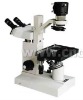
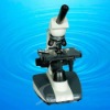
| Biological Microscope 107-E |
|
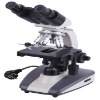
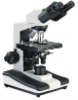
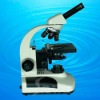
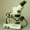
| 107-E Biological Microscope |
|
OUTFIT | MODEL | | 107E | | Achromatic Objectives | 4X,10X,40X ( S ).100X( S, Oil ) | - | | Eyepiece | WF10X,(WF16X for choice) | - | Eyepiece head | Monocular head | | | Compensation Free Binocular Head | - | | Compensation Free Binocular Head | | | Nosepiece | Quadruple | - | | Stage | Double Layer Mechanical Stage 115x125mm,Moving Range 75x45mm | - | | Focusing | Coaxial Coarse and Fine Adjustment, Focusing Range 30mm, Focusing Interval 0.002mm | - | Illumination, condenser
NA=1.25 | 6V/20W Halogen Lamp, Adjustable Brightness | - | | Power | AC Input 220V or 110V, DC Output 6V | - |
|
Microscope is an instrument to see objects too small for the naked eye. The science of investigating small objects using such an instrument is called microscopy. Microscopic means invisible to the eye unless aided by a microscope.
There are many types of microscopes, the most common and first to be invented is the optical microscope which uses light to image the sample. Other major types of microscopes are the electron microscope (both the transmission electron microscope and the scanning electron microscope) and the various types of scanning probe microscope.
Microscopes can be separated into several different classes. One grouping is based on what interacts with the sample to generate the image, i.e., light (optical microscopes), electrons (electron microscopes) or a probe (scanning probe microscopes). Alternatively microscopes can be classed on whether they analyse the sample via a scanning point (confocal optical microscopes, scanning electron microscopes and scanning probe microscopes) or analyse the sample all at once (wide field optical microscope and transmission electron microscopes).
The wide field optical microscope and transmission electron microscope use the theory of lenses (optics for light microscopes and electromagnet lenses for electron microscopes) in order to magnify the image generated by the passage of a wave through the sample, or reflected by the sample. The waves used are electromagnetic (in optical microscopes) or electron beams (in electron microscopes). Resolution in these microscopes is limited by the wavelength of the radiation used to image the sample, shorter wavelengths allow a higher resolution.
Scanning optical and electron microscopes, like the confocal microscope and scanning electron microscope, use lenses to focus a spot of light/electrons onto the sample then analyse the reflected and/or transmitted waves. The point is then scanned over the sample to analyse a rectangular region. Magnification of the image is achieved by displaying the data from scanning a small sample area on a large screen. These microscopes have the same resolution limit as wide field optical and electron microscopes.
Scanning probe microscopes also analyse a single point in the sample and then scan the probe over a rectangular sample region to build up an image. As these microscopes do not use electromagnetic or electron radiation for imaging they are not subject to the same resolution limit as the optical and electron microscopes described above.
The most common type of microscope—and the first invented—is the optical microscope. This is an optical instrument containing one or more lenses producing an enlarged image of an sample placed in the focal plane. Optical microscopes have refractive glass and occasionally of plastic or quartz, to focus light into the eye or another light detector. Mirror-based optical microscopes operate in the same manner. Typical magnification of a light microscope, assuming visible range light, is up to 1500x with a theoretical resolution limit of around 0.2 micrometres or 200 nanometers. Specialized techniques (e.g., scanning confocal microscopy, Vertico SMI) may exceed this magnification but the resolution is diffraction limited. The use of shorter wavelengths of light, such as the ultraviolet, is one way to improve the spatial resolution of the optical microscope, as are devices such as the near-field scanning optical microscope.
Sarfus, a recent optical technique increases the sensitivity of standard optical microscope to a point it becomes possible to directly visualize nanometric films (down to 0.3 nanometer) and isolated nano-objects (down to 2 nm-diameter). The technique is based on the use of non-reflecting substrates for cross-polarized reflected light microscopy.
Ultraviolet light enables the resolution of microscopic features, as well as to image samples that are transparent to the eye. Near infrared light images circuitry embedded in bonded silicon devices, as silicon is transparent in this region. Many wavelengths of light, ranging from the ultraviolet to the visible are used to excite fluorescence emission from objects for viewing by eye or with sensitive cameras.
Phase contrast microscopy is an optical microscopy illumination technique in which small phase shifts in the light passing through a transparent specimen are converted into amplitude or contrast changes in the image. A phase contrast microscope does not require staining to view the slide. This microscope made it possible to study the cell cycle.
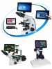
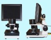
The traditional optical microscope has recently been modified into a digital microscope, where, instead of directly viewing the object, a charge-coupled device (CCD) is used to record the image, which is then displayed on a computer monitor.